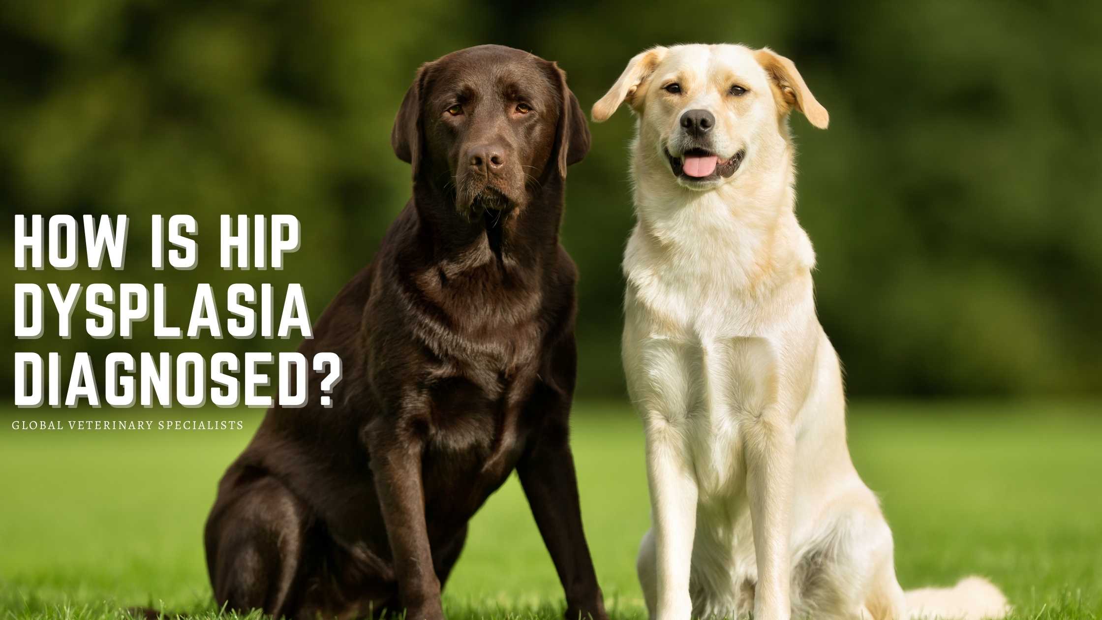
Hip dysplasia (HD) is a condition that results from the abnormal development of the hip joint in which the ball does not fit snugly into the socket. It can affect both cats and dogs of any size or breed. A comprehensive examination must be completed to rule out other causes of hind lameness or conditions that could exacerbate HD. Radiographs are then obtained to confirm the diagnosis of hip dysplasia.
Other causes of hind lameness include but may not be limited to the following:
- Hip luxation
- Patella luxation
- Joint infection
- Spinal or neurological abnormalities
- Fractures of the femoral head (the ball)
- Trauma or injury to other soft tissue limb structures
- Orthopedic conditions associated with developmental growth such as osteochondritis dessicans (OCD), panosteitis, or Legg-Perthes disease
The veterinarian will review the patient’s medical history and, if available, genetic background. It is essential to relay information about your pet’s nutrition, symptoms, activity level, and prescribed medications. They will also perform both a physical and orthopedic examination.
During the orthopedic exam, the dog’s posture and ambulation are observed, and palpation is performed on all four limbs. The latter focuses on each long bone, the major joints, muscle mass, the spine, and the supporting soft tissues. The hip joint is usually examined last to rule out other causes of pain and inflammation.
Excessive joint laxity can be palpated using a diagnostic test, called the Ortolani test, in dogs who do not resist manipulation. An Ortolani sign is beneficial when grading young dogs with HD, particularly in puppies up to about eight to ten weeks, as mild laxity is normal. The Ortolani sign is an indication of hip dysplasia in dogs older than six months.
Imaging Evaluation
Because of the importance of obtaining radiographs to make an accurate diagnosis, it may be best to sedate overly excited dogs or those experiencing pain. That will allow for perfectly positioned imaging to be completed with minimal X-ray exposure. Two views, ventral-dorsal and lateral, are usually sufficient in the diagnosis of this condition.
Radiographic indications of HD include evidence that the femoral head is not seated deeply into the acetabulum – approximately 50 percent of the head should be superimposed inside the socket – and the absence of joint incongruity. Secondary osteoarthritis may or not be present and to a variable degree of severity.
The severity of the laxity and, if present, arthritis must be determined by obtaining views of both hips. The combination of both the clinical and radiographic findings is essential for selecting the best treatment options. A total hip replacement (THR) is often recommended in animals with moderate to severe HD.
What Are the Signs of Hip Dysplasia?
Common symptoms include:
- Limping
- Decreased activity
- A “bunny hop” gait
- Difficulty rising after rest
- Hesitance to jump or climb stairs
- Premature tiring, especially during exercise
- Weight shifting from one or both hind feet
- Slightly arched back, resulting from weight shifting
Schedule an Exam Appointment Today
Global Veterinary Specialists recognize that dogs and cats encounter orthopedic injuries, disabling difficulties, and diseases that can impact their quality of life and are committed to restoring mobility by treating hip dysplasia and other complex problems. Each GVS surgeon has more than twenty years of veterinary and surgical experience.
We are teachers, mentors, inventors, clinical researchers, and veterinary surgeons driven to achieve excellent results for every animal we see regardless of their age or breed. Please contact us today to learn about the various options for treating hip dysplasia and how we can help restore your pet’s quality of life.